
 The Radiology department at Healthcare has a state of art imaging modalities under one roof. Investment in state-of-the-art technology in collaboration with leading manufacturers like GE, Sonosite, Electro Medicals helps the patient get high resolution, excellent quality reports ensuring a quick diagnosis of the disease.
Services Provided :
- Digital Sonography
- Digital 3D Sonography
- Digital Color Doppler Study
- Hi-Power 300 MA capacity X-Ray
- X-Ray procedures – HSG, IVP, and Barium Meal
- Mammography
- Bone Mineral Densitometry
Sonography
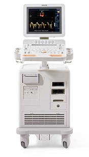
Sonography is a non invasive imaging modality using sound waves and is extremely safe even in pregnancy as it does not involve any radiation. The machine installed in the centre is DIGITAL ULTRASONOGRAPHY GE LOGIC PRO 3i
New sonography machine installed : Philips HD-7 [3-D]Series Sono/Color Doppler/Echocardiography machine.
Sonography has applications for the entire body from head to toe. Male or female, from infant to adult, throughout your lifetime, Sonography can play an important role in your healthcare. Indeed, versatile, safe, non-invasive and yet effective, the importance of ultrasonography in medicine cannot be underplayed.
Sonography, or ultrasound, utilizes high frequency sound waves (not x-rays) to obtain diagnostic images. Ultrasound imaging is used to evaluate many parts of the body, including the abdomen, blood vessels, fetus of pregnant women, superficial body structures, and newborn brain to name only a few.
 Ultrasonography enables to detect and investigate :
- All diseases of the organs of the abdominal cavity in early stages.
- Tumors of uterus and ovaries and abnormalities of reproductive organs.
- Maturation of eggs and changes of endometrium in different stages of menstrual cycle (early pregnancy, including entopic pregnancy).
- Development of fetuses and possible malformations of fetuses.
- Position of the fetus, position of the placenta in the uterus and changes in it. It is also possible to estimate the quantity of amniotic fluid, evaluate heart function and breathing movements of the fetus.
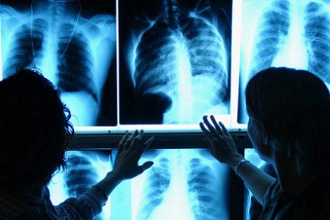
Digital X-RAY
Digital X-rays are a form of electromagnetic radiation, just like visible light. A computer or special film is used to record the images that are created. At healthcare Centre, X-rays are taken on DIGITAL X-RAY Konica Minolta Made in Japan, which provides superior and high quality x-rays. The centre also has a portable x-ray machine from Meditronix systems which has an image intensification system. There is no discomfort from x-ray exposure. Patients may be asked to stay still in a particular positions for a short period of time.
Preparations for Digital X-Ray
Inform the health care provider prior to the examination if you are pregnant or have an IUD inserted. You will be asked to remove all jewellery and wear a center gown during the digital x-ray examination because metal and certain clothing can obscure the images and require repeat studies.
Colour Doppler
Color Doppler is color-encoded Sonography of the blood vessels. It is like Sonography and there is no involvement of any injection of contrast. It complements 2-D echocardiography and conventional Doppler techniques by providing color flow maps that improve the spatial characterization of flow disturbances.
Patient Benefits :
- Faster Examination
- High resolution images for detection of subtle abnormalities.
- Vascular information
 Mammography
A mammogram is an x-ray examination of the breasts, used to detect and diagnose breast diseases. Mammography is the most effective method of detecting cancer at an early stage, before the woman or a physician can feel it. Screening mammography is used as a preventive measure for women who have no symptoms of breast disease. A screening mammogram usually involves two views of each breast. The American Cancer Society recommends that all women aged 40 and over have a screening mammogram every year as part of a breast health program, which also includes an annual breast examination by a healthcare professional.
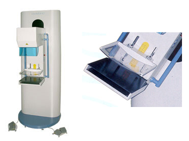
At HEALTHCARE mammography is done by VISIMAM 501.
VISIMAM is a state –of –the-art breast cancer detection system that is design to improve the quality and comfort of mammograms. The system produces high quality of images that allow for better visualization of breast tissue, aiding the early detection of breast cancer. This advanced system also includes features, that reduce procedure times and improve patient comfort. VISIMAM series of Mammography Systems delivers high quality breast images-consistently, efficiently and easily.
Bone Mineral Densitometry
 A bone mineral density (BMD) test measures the density of minerals (such as calcium) in your bones to estimate the strength of your bones. We all lose some bone mass as we age. Bones naturally become thinner (called osteopenia) as you grow older, because existing bone is broken down faster than new bone is made. As this occurs, our bones lose calcium and other minerals and become lighter, less dense, and more porous. This makes the bones weaker and increases the chance that they might break (fracture).
With further bone loss, osteopenia leads to osteoporosis . So the thicker your bones are, the longer it takes to get osteoporosis. If your bone density is lower than normal, you can take steps to increase your bone strength and reduce your chances of having a fracture. Some ways to increase bone density and strength include combining calcium and vitamin D supplements with weight-bearing exercise (such as walking), weight training (such as lifting weights or using weight machines), and using medicines.
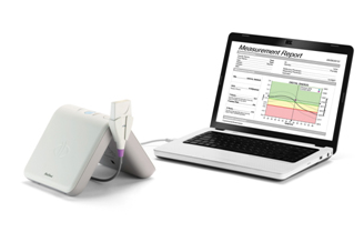
At HEALTHCARE we have installed MiniOmni ( Ultrasound Based BMD) machine which is quick, painless, and does not use potentially harmful radiation like X-rays. Osteoporosis needs to be diagnosed accurately and precisely, and its progress monitored over a period of time. The simplest solution for this purpose is the use of ultrasound for measurement of speed of sound in the bones.